The Revolution in Digital Fluoroscopy Systems: Advantages of Digital Flat Panel Detectors
 |
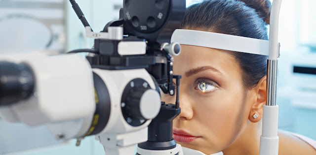
| Digital Fluoroscopy Systems |
Digital fluoroscopy systems is a type of medical imaging that shows a continuous X-ray image on a monitor, much like an X-ray movie. It is primarily used in image-guided procedures to capture real-time moving images of internal organs, bones, blood vessels, and other tissues. Some of the key areas where fluoroscopy is used include cardiology, radiology, orthopaedics, gastroenterology and vascular surgery. Fluoroscopy has been around since the late 1890s as a useful imaging tool to guide minimally invasive procedures. Traditionally, fluoroscopy systems used image intensifier-based technology and analogue imaging. However, over the past decade there has been a digital transformation of fluoroscopy with the introduction of digital flat panel detectors.
Advantages of Digital Flat Panel Detectors
The biggest difference between traditional image intensifier-based fluoroscopy and modern digital flat panel fluoroscopy systems is the detector technology. Traditional systems used a vacuum tube image intensifier to convert X-rays into a visible light image, which was then recorded on film or video camera. Digital flat panel detectors directly convert X-rays into digital signals without the need for an intermediate light conversion step. This provides several key advantages over traditional technology:
- Image quality: Digital detectors provide higher resolution images compared to analogue systems. They have less optical distortion, better low contrast resolution and less noise which results in sharper images.
- Dose reduction: Digital Fluoroscopy System require much lower radiation doses compared to traditional systems as they have higher detection efficiency. Studies show radiation dose reduction up to 70-80% with digital detectors. This is hugely beneficial for patients and operators.
- Archiving and sharing: Digital images can be stored, retrieved and electronically shared much more easily compared to physical films. This improves workflow and enables remote consultation.
- Advanced imaging features: Digital detectors support powerful features like fluoro-motion, roadmapping, digital subtraction, tomosynthesis etc which provide additional clinical benefits.
Applications of Digital Fluoroscopy
With the advantages outlined above, digital fluoroscopy systems technology is transforming minimally invasive procedures across multiple clinical specialties:
Cardiology: To guide coronary angioplasty and stent placement, ablation procedures for arrhythmias with live imaging of catheter movement. Roadmapping and digital subtraction help optimize visualization.
Radiology: Used for image-guided biopsies, drain placements, vertebroplasty, kyphoplasty with high quality real-time imaging and minimal dose.
Orthopaedics: Enables minimally invasive joint replacements and fracture care with intra-operative imaging feedback. Provides live bone visualization during surgical navigation.
Gastroenterology: Essential for ERCP, stent placements, Heller's myotomy etc to guide wire and catheter movement in real-time imaging of bile ducts, pancreas and esophagus anatomy.
Vascular/Interventional Radiology: Critical for angiography, angiogram, angioplasty, stent placements, thrombolysis with clear visualization of vessels, tumors and thrombi in motion permitting accurate catheter navigation to target sites.
Urology: Facilitates PCNL (percutaneous nephrolithotomy) for kidney stone removal and other procedures with real-time imaging of urinary tract. Cyclo ablation for prostate cancer uses image-guided navigation.
Emerging Areas of Focus
As digital fluoroscopy systems technology reaches a mature phase, vendors and researchers are exploring new enhancements and additional applications:
- Integrated surgical navigation: Combining high definition 3D/4D imaging, patient tracking, instrument tracking for image-guided minimally invasive surgeries with live cross-sectional and projection views.
- Artificial intelligence: Use of AI for automated image enhancement, dose control, procedural analysis, surgeon guidance and predictive diagnostics based on large datasets.
- Imaging fusion: Fusing digital fluoroscopy systems with pre-operative CT/MRI for improved anatomical context during procedures through image registration techniques.
- Low dose systems: Continued optimization of detectors, hardware and software protocols is lowering patient exposure further while maintaining excellent image quality.
- Extended clinical benefits: Novel applications like chronic pain management, nerve ablation, pediatric imaging etc. are further expanding fluoroscopy's clinical utility and streamlining patient care workflows.
The digital transformation of fluoroscopy with flat panel detectors is a key technological innovation driving minimally invasive procedures across major clinical specialties. The improvements in image quality, dose reduction capabilities and flexible advanced features have positioned digital fluoroscopy systems as an indispensable tool for image-guided interventions. Going forward, continued enhancements through integration with AI, augmented/virtual reality and surgical robots hold promise to take fluoroscopy assisted interventions to even higher levels of precision and safety
Get more insights on Digital Fluoroscopy System
About Author:
Vaagisha brings over three years of expertise as a content editor in the market research domain. Originally a creative writer, she discovered her passion for editing, combining her flair for writing with a meticulous eye for detail. Her ability to craft and refine compelling content makes her an invaluable asset in delivering polished and engaging write-ups.
(LinkedIn: https://www.linkedin.com/in/vaagisha-singh-8080b91)

%20Filter%20Market.jpg)
Comments
Post a Comment